


Transkranielle Ultraschalldiagnostik
English version below
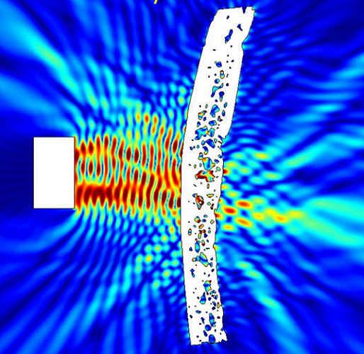
Ultraschall ist das am weitesten verbreitete bildgebende Verfahren in der Medizin. Jedoch stört der Schädelknochen seine Anwendung in der Diagnostik und Therapie des Hirns häufig unkontrollierbar. Ziel des Projekts „Transkranielle Ultraschalldiagnostik“ ist es, Indikationen des diagnostischen Ultraschalls durch präzise Vermessung und Korrektur der Schallausbreitung im Knochen erweitern zu können.
An der Zahl der Prozeduren gemessen ist Ultraschall in der Medizin das am weitesten verbreitete bildgebende Verfahren. Jenseits der Radiologie setzen es praktisch alle Fachdisziplinen zur Diagnostik ein. Vorteil einer Ultraschall-Untersuchung ist: Im Gegensatz zu Computertomografie und Magnetresonanztomografie kann sie am Patientenbett durchgeführt werden. In den letzten Jahren haben sich zudem ultraschallgestützte therapeutische Verfahren entwickelt. Ein Schwerpunkt liegt dabei in der Therapie neurologischer Indikationen (z.B. thermische Ablationen im Hirn bei essentiellem Tremor, Parkinson oder Epilepsie; Neuromodulation). Die Anwendung von Ultraschall in Diagnostik und Therapie des Hirns wird jedoch erschwert: Der Schädelknochen stört häufig die Schallausbreitung unkontrollierbar.
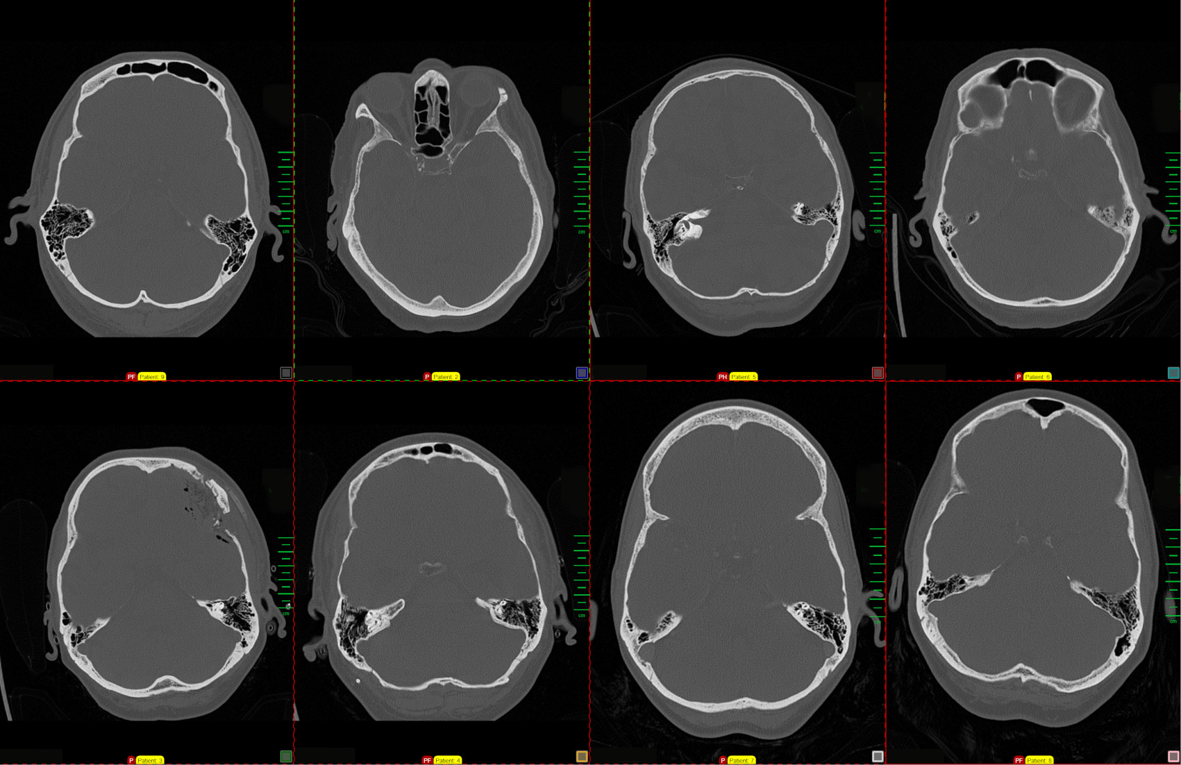
Basierend auf CT- und Ultraschalldatensätzen des Klinikums Nürnberg will das Projektteam diese Störungen verstehen und technische Verfahren zur Korrektur aufzeigen. Dadurch würden die diagnostischen wie therapeutischen Prozeduren erleichtert. Neben bereits existierenden therapeutischen Einsätzen des Ultraschalls z.B. im Rahmen einer Gewebeablation ist der diagnostische Einsatz interessant. Dieser ist aktuell nur bei Kleinkindern durch noch nicht verkalkte Fontanellen möglich bzw. bei der transkraniellen Doppler-Sonographie durch kleine Schallfenster. Ziel wäre eine Erweiterung der Indikationen des diagnostischen Ultraschalls (z.B. Blutungslokalisation, Mittellinienverlagerung, verbesserte Darstellung der Blutgefäße) über die bekannten und oft unzureichenden Schallfenster hinaus.
Transcranial Ultrasound Diagnostics
Aberration of ultrasound propagation in the skull bone complicates diagnosis and therapy of brain diseases. The project „Transcranial Ultrasound Diagnostics“ aims to develop a method for measuring and correcting these aberrations.
Ultrasound is by far the most widely used imaging modality in medicine. However, cranial bone often interferes uncontrollably with sound propagation in diagnostics and therapy of the brain. Aim of the project „Transcranial Ultrasound Diagnostics“ is to expand indications of ultrasound by precise measurement and correction of sound propagation and aberration in bone.
In terms of the number of procedures, ultrasound is the most widely used imaging modality in medicine. Beyond radiology, virtually all medical disciplines use it for diagnostics. Unlike computer tomography and magnetic resonance imaging, ultrasound imaging can be performed at the point of care. In recent years, a plurality of ultrasound-based therapeutic procedures have been developed. One focus is on the therapy of neurological indications (e.g., thermal ablations in the brain for essential tremor, Parkinson’s disease or epilepsy; neuromodulation for stimulating a reversible neural response). They are well tolerated by patients and some have already gained clinical approval. However, the use of ultrasound in the brain is difficult: skull bone interferes a priori uncontrollably with the propagation of sound.
Based on CT and ultrasound datasets from Klinikum Nuremberg, the project team want to understand ultrasound aberrations and identify technical procedures to correct them. In addition to the mentioned therapies, diagnostic ultrasound in the brain is of particular interest. Currently imaging is only possible in small infants through not yet calcified fontanelles, or in transcranial Doppler sonography through small sound windows. It is the goal to eventually expand indications of diagnostic ultrasound (e.g., hemorrhage localization, midline shift, improved visualization of blood vessels) beyond the known and often inadequate sonic windows.
Projektbeteiligte:
-
Prof. Dr. Florian Steinmeyer
Technische Hochschule Nürnberg, AMP, Angewandte Mathematik, Physik und Allgemeinwissenschaften
-
Milan Fritsche
Technische Hochschule Nürnberg, AMP, Angewandte Mathematik, Physik und Allgemeinwissenschaften
-
Prof. Dr. med. Michael Lell
Klinikum Nürnberg, Institut für Radiologie und Nuklearmedizin
-
Dr. Josefin Ammon
Klinikum Nürnberg, Institut für Medizinische Physik
-
Dr. Barbara Schmid
Klinikum Nürnberg, Abteilung für Klinische Neurophysiologie